
 By Cindy Adoptante
Get 11 Sample Recipes from Our Bestselling Mediterranean Cookbook.
Get our 11 Sample Recipes from our Best Selling Mediterranean Protein Friendly Cookbook. These are the most delicious recipes our retreat guests love the most. Easy, simple with few ingredients per meal. Make delicious heart-friendly and healthy meals you enjoy every day.
Xanthelasma (Cholesterol Spots on Eyelids): What Those Yellow Patches Are Telling You
Somewhere in her late forties or early fifties, a woman looks in the mirror and notices something new near the inner corners of her eyes: a soft, flat, faintly yellow patch, symmetrical on both sides, that was not there a year ago. It does not hurt. It does not itch. It just sits there, slightly raised, slightly waxy-looking, and it raises an obvious question. What is this, and should I be worried about it?
The patch has a name: xanthelasma. It is one of the more common visible skin findings in midlife, and it is also one of the more medically interesting ones, because unlike most cosmetic skin changes, xanthelasma has a genuine, well-documented connection to cardiovascular health, one that a large Scandinavian study eventually traced with more precision than almost any other visible physical marker researchers have studied. That connection is real, but it is also more nuanced than most headlines about it suggest, and the timing of when xanthelasma tends to appear, right around the menopause transition, is not a coincidence.
This article walks through exactly what xanthelasma is, why it forms, what the research shows about its connection to heart disease, why it shows up so often in midlife women specifically, what the honest treatment picture looks like, and how it connects to the broader nutrition picture that matters most during this life stage.
What Is Xanthelasma?
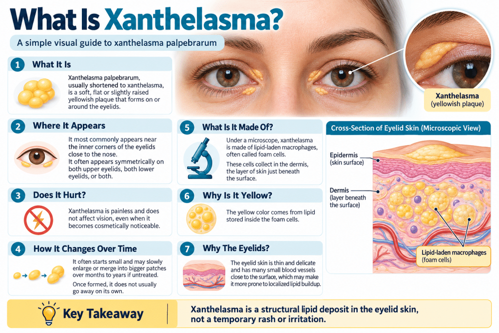
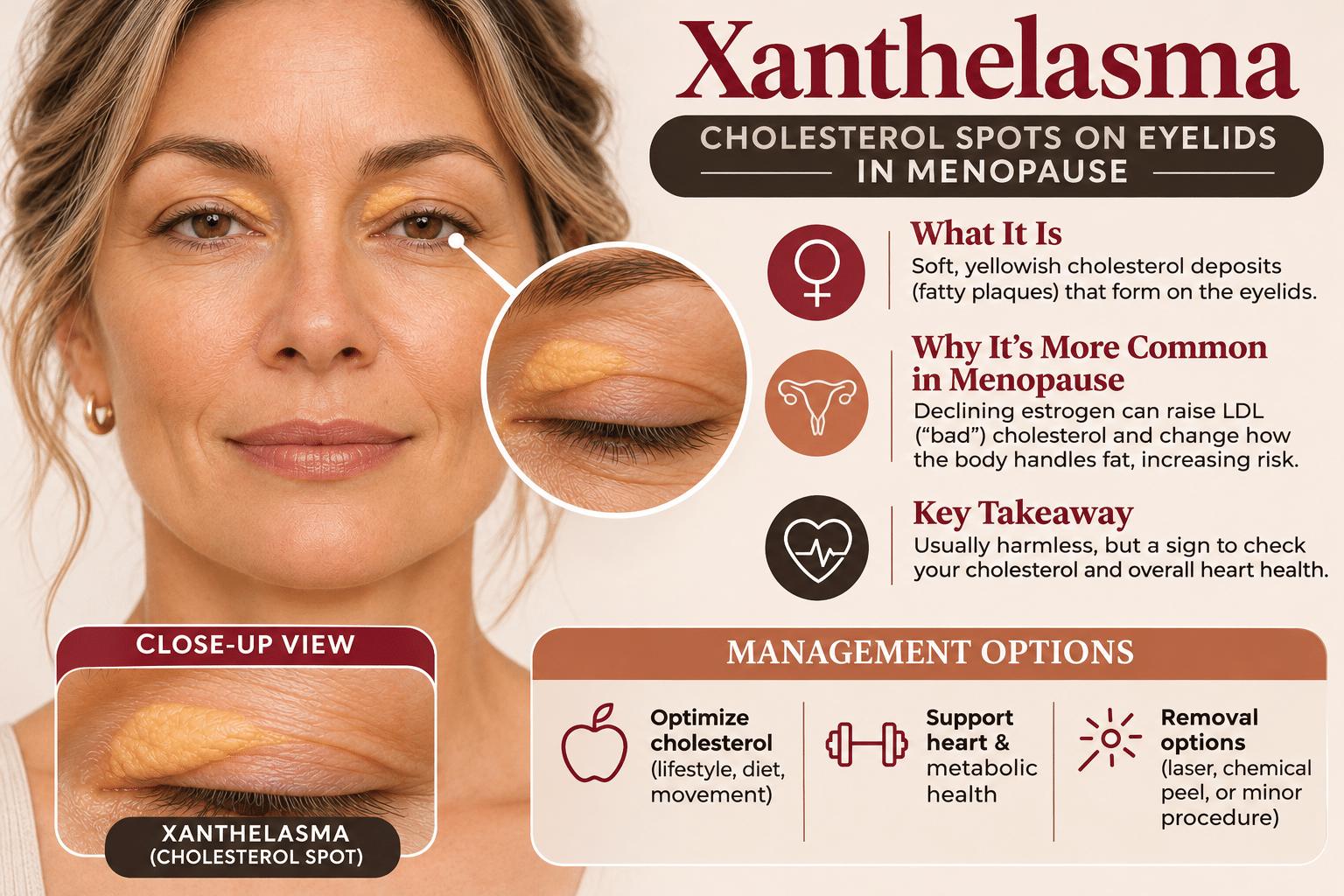
Xanthelasma palpebrarum, usually shortened to xanthelasma, is a soft, flat or slightly raised, yellowish plaque that forms on or around the eyelids, most often at the inner corners near the nose. It usually appears on both sides, on the upper lids, lower lids, or both, though it can appear on just one side.
Key features:
- Soft, flat or slightly raised
- Yellow or orange-yellow color
- Most often at the inner corners of the eyelids
- Painless, doesn’t itch, and doesn’t affect vision
- Starts small and can slowly grow or merge into larger patches
- Doesn’t go away on its own
What it’s made of: Under a microscope, xanthelasma is made of lipid-laden immune cells called macrophages. These cells take up so much cholesterol that they look foamy, earning the name “foam cells.” They collect in the dermis, the layer just beneath the skin’s surface, and their fat content gives the patch its yellow color.
The eyelid’s thin, delicate skin and dense network of small blood vessels may make it especially prone to these deposits.
What Is the Difference Between Xanthelasma and Corneal Arcus?
Xanthelasma is often mentioned alongside a second, distinct eye-area finding called corneal arcus, sometimes called arcus senilis or arcus corneae. Corneal arcus is a grayish-white ring or arc that forms around the outer edge of the cornea, the clear front surface of the eye, rather than on the eyelid skin itself. Like xanthelasma, it results from lipid deposition, in this case in the peripheral cornea rather than the dermis of the eyelid.
The two conditions are related in mechanism, both involve lipid deposits in tissue around the eye, and they are frequently discussed together in the medical literature, but they are anatomically distinct findings, appear in different tissues, and, as the research in Section 7 of this article lays out in detail, do not carry identical evidence for cardiovascular risk.
Corneal arcus is common enough in older adults that it is sometimes considered a near-universal feature of aging past a certain point, which is part of why its individual predictive value is weaker and more debated than xanthelasma’s. Because corneal arcus deserves its own full treatment rather than a rushed summary, this article touches on it only where the research specifically requires that comparison, and treats it as a related topic worth exploring on its own in future content.
Get 11 Sample Recipes from Our Bestselling Mediterranean Cookbook.
Get our 11 Sample Recipes from our Best Selling Mediterranean Protein Friendly Cookbook. These are the most delicious recipes our retreat guests love the most. Easy, simple with few ingredients per meal. Make delicious heart-friendly and healthy meals you enjoy every day.
What Causes Xanthelasma?
The core mechanism behind xanthelasma is lipid accumulation inside dermal macrophages, but several distinct pathways can lead to that outcome:
- Elevated LDL cholesterol: In a substantial portion of cases, xanthelasma is associated with elevated low-density lipoprotein (LDL) cholesterol, the form of cholesterol most strongly tied to atherosclerotic cardiovascular disease. Excess circulating LDL increases the amount of lipid material available to be taken up by macrophages in the skin.
- Genetic lipid disorders: Some people with xanthelasma have an underlying inherited condition affecting lipid metabolism, such as familial hypercholesterolemia or familial dysbetalipoproteinemia, in which cholesterol and other lipids run persistently high regardless of diet. In these cases, xanthelasma can be one of the earliest visible clues to an underlying genetic condition that would otherwise go undetected without bloodwork.
- Age-related changes in lipid clearance: Independent of any diagnosed lipid disorder, the body’s efficiency at clearing LDL cholesterol from the bloodstream tends to decline with age, contributing to a general rise in circulating lipid levels that can make dermal deposition more likely over time.
- Lipid peroxidation, even without elevated cholesterol: This is the least well-known and most interesting piece of the puzzle, covered in detail in the next section: a meaningful share of people with xanthelasma have completely normal standard cholesterol panels.
- Family history: Xanthelasma appears to run in families independent of diagnosed lipid disorders, suggesting a heritable component to who develops the finding beyond cholesterol numbers alone.
Why Can Xanthelasma Appear With Normal Cholesterol?
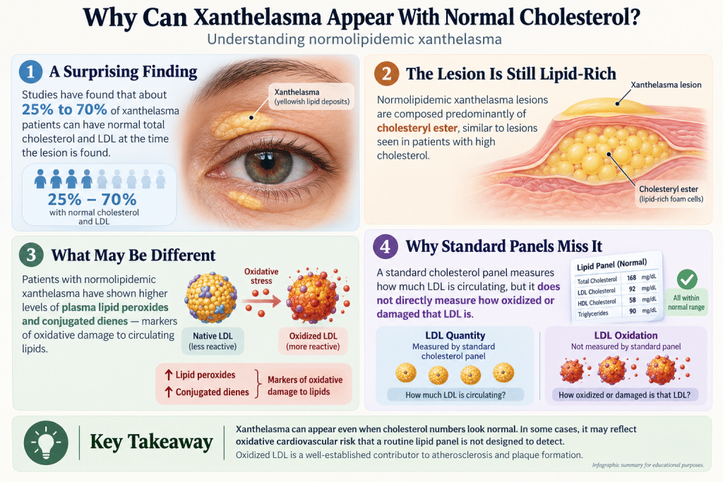
One of the more counterintuitive findings in the xanthelasma research is how common it is for the lesion to appear in people whose standard cholesterol panel comes back entirely normal. Research on this specific subgroup, often referred to as normolipidemic xanthelasma, has found that roughly a quarter to as much as seventy percent of xanthelasma patients across different study populations have normal total cholesterol and LDL levels at the time the lesion is identified.
This does not mean the finding is meaningless in these patients. Studies specifically analyzing normolipidemic xanthelasma have found that the lesions themselves are composed predominantly of cholesteryl ester, similar in composition to lesions in patients with elevated cholesterol, rather than showing a fundamentally different lipid profile. More notably, patients with normolipidemic xanthelasma have been shown to have significantly higher levels of plasma lipid peroxides and conjugated dienes, markers of oxidative damage to circulating lipids, compared to matched controls without the lesion.
The practical implication is worth sitting with: a standard cholesterol panel measures the total quantity of LDL circulating in the blood, but it does not directly measure how oxidized or damaged that LDL is. Oxidized LDL is itself a well-established contributor to atherosclerosis, arguably a more direct driver of plaque formation than raw LDL quantity alone. Xanthelasma appearing in someone with a “normal” cholesterol number may, in at least some cases, reflect this oxidative dimension of cardiovascular risk that a standard lipid panel simply is not designed to catch.
“This is the piece of the research that changed how I think about xanthelasma entirely. I used to assume, like most people probably do, that a clean cholesterol panel meant a visible marker like this was ‘just cosmetic.’ The oxidative stress angle tells a more complete story. It is part of why I care so much about the quality of fat and the antioxidant density of what we eat, not just the numbers on a standard lab panel, because the numbers alone do not always capture what is happening at the cellular level. I am not a physician, and this is not a diagnosis of anyone’s individual risk, but it is exactly the kind of nuance that makes nutrition, not just supplementation, the more powerful lever.”
Terry Tateossian, Founder of The House of Rose
Get 11 Sample Recipes from Our Bestselling Mediterranean Cookbook.
Get our 11 Sample Recipes from our Best Selling Mediterranean Protein Friendly Cookbook. These are the most delicious recipes our retreat guests love the most. Easy, simple with few ingredients per meal. Make delicious heart-friendly and healthy meals you enjoy every day.
Why Does Xanthelasma Show Up After Menopause?
Xanthelasma prevalence rises with age in both sexes, but it shows a particularly notable jump in women around the menopause transition, and the reason comes down to a well-documented shift in lipid metabolism driven by declining estrogen.
Research comparing postmenopausal women to age-matched premenopausal women has found meaningful, measurable increases in the lipid markers most relevant to xanthelasma formation: total cholesterol roughly 10 percent higher, LDL cholesterol roughly 14 percent higher, and apolipoprotein B, a marker that reflects the total number of LDL and other atherogenic particles circulating in the blood, roughly 8 percent higher in postmenopausal women, even after adjusting for age.
The mechanism behind this shift centers on estrogen’s role in LDL clearance. Estrogen supports the activity of LDL receptors, the structures on liver cells responsible for pulling LDL cholesterol out of the bloodstream and clearing it from circulation. As estrogen declines through perimenopause and into postmenopause, LDL receptor efficiency drops with it, and LDL cholesterol accumulates in the blood at a higher baseline level than it did premenopausally, independent of any change in diet or body weight.
Research tracking these changes has found they occur predominantly in a relatively compressed window around late perimenopause and early postmenopause, rather than gradually across decades, which lines up with the clinical observation that xanthelasma often seems to “appear out of nowhere” in a woman’s late forties or fifties.
This is precisely why xanthelasma is a meaningful marker to flag for THOR’s audience specifically. It is not simply a skin finding of aging in general; it is, for a substantial number of women, a visible expression of a specific, well-characterized hormonal shift in lipid metabolism that happens to coincide almost exactly with the menopause transition.
What Did the Copenhagen City Heart Study Find?
The single most important piece of research connecting xanthelasma to cardiovascular outcomes comes from the Copenhagen City Heart Study, published in the BMJ in 2011 by Christoffersen and colleagues. This was a large, long-running prospective cohort study, one of the most methodologically rigorous designs available in observational research, and it is worth understanding exactly what it did and exactly what it found, because the finding gets oversimplified constantly in popular coverage.
The Study Design
Researchers followed 12,745 people between the ages of 20 and 93, all free of known ischemic vascular disease at the start of the study, from 1976 through 1978, with follow-up continuing until May 2009. Follow-up completeness was total: 100 percent of participants were tracked for the full study period, an unusually strong feature for a study spanning more than three decades.
Baseline Prevalence
At the start of the study, 4.4 percent of participants had xanthelasma and 24.8 percent had corneal arcus, giving researchers a large enough sample of both findings to analyze their outcomes with statistical confidence.
The Xanthelasma Findings
After adjusting for age, sex, and the standard, well-established cardiovascular risk factors (things like blood pressure, smoking status, diabetes, and cholesterol levels), the presence of xanthelasma at baseline was associated with a meaningfully increased risk of several cardiovascular outcomes over the following decades: a hazard ratio of 1.48 for myocardial infarction (heart attack), 1.39 for ischemic heart disease more broadly, and 1.14 for death from any cause.
In practical terms, this means people with xanthelasma had roughly 48 percent higher risk of heart attack and 39 percent higher risk of ischemic heart disease than people without it, even after statistically accounting for their cholesterol levels and other standard risk factors. That last detail is the single most important finding in the entire study: the elevated risk persisted independent of the person’s measured cholesterol, meaning xanthelasma appears to be flagging something about cardiovascular risk that a standard lipid panel does not fully capture, consistent with the oxidative-stress mechanism discussed in Section 4.
What Xanthelasma Was Not Associated With
The same analysis found hazard ratios of 0.94 for ischemic stroke and 0.91 for ischemic cerebrovascular disease broadly, close to no association, and in fact slightly protective point estimates, though these were not statistically significant findings in either direction. The elevated risk found in this study was specific to coronary and cardiac outcomes, not a general elevation in all vascular disease risk.
This is one of the largest and longest-running studies ever conducted on a visible physical marker of cardiovascular risk, and the finding that xanthelasma predicts heart attack risk independent of cholesterol level is truly significant, not overstated marketing language. It is also, importantly, not a diagnosis or a guarantee. It is a population-level statistical association from a single (if very strong) study, and it should be understood the same way any single risk marker should: as one data point worth taking seriously, not as a crystal ball.
Get 11 Sample Recipes from Our Bestselling Mediterranean Cookbook.
Get our 11 Sample Recipes from our Best Selling Mediterranean Protein Friendly Cookbook. These are the most delicious recipes our retreat guests love the most. Easy, simple with few ingredients per meal. Make delicious heart-friendly and healthy meals you enjoy every day.
What Did the Study Not Show About Corneal Arcus?
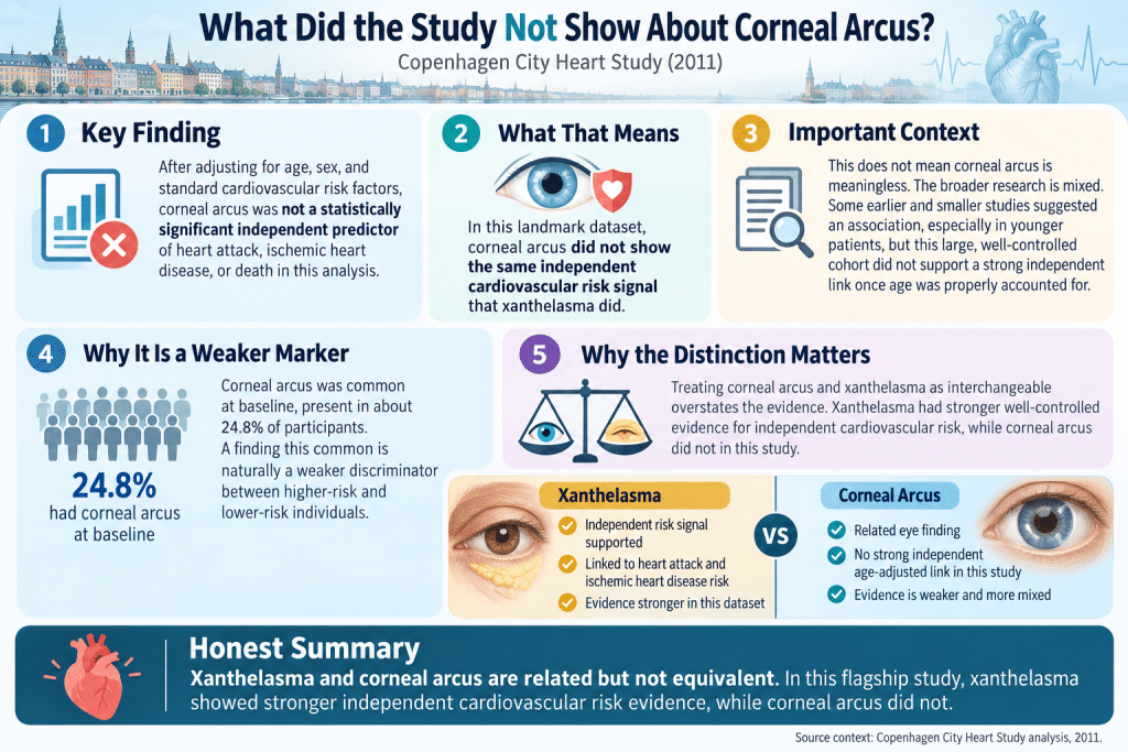
Because xanthelasma and corneal arcus are so often mentioned in the same breath, it would be easy to assume the Copenhagen City Heart Study found the same elevated cardiovascular risk for both. It did not, and this distinction matters enough to spell out clearly.
In the same 2011 analysis, after the same adjustment for age, sex, and standard cardiovascular risk factors, the hazard ratios calculated for corneal arcus and cardiovascular outcomes did not reach statistical significance. In other words, once age and the standard risk factors were properly accounted for, this particular study did not find good evidence that corneal arcus independently predicts heart attack, ischemic heart disease, or death in the way xanthelasma did.
This does not mean corneal arcus is meaningless, and it does not mean no other study has ever found an association; the broader literature on corneal arcus is more mixed than the literature on xanthelasma, with some earlier and smaller studies suggesting an association, particularly in younger patients, while others, including this large, well-controlled cohort, do not support a strong independent link once age is properly controlled for.
Corneal arcus is also simply far more common than xanthelasma, present in roughly a quarter of this study’s participants at baseline, which naturally makes it a weaker discriminator between higher-risk and lower-risk individuals; a marker present in one in four people at baseline cannot function as a highly specific single risk indicator.
The honest summary: xanthelasma has strong, well-controlled, specific evidence connecting it to elevated heart attack and ischemic heart disease risk independent of cholesterol level. Corneal arcus is a related finding, worth understanding in its own right, but the flagship evidence connecting it to cardiovascular risk independent of age is considerably weaker, at least in this landmark dataset.
Treating the two as interchangeable overstates what the research on corneal arcus specifically shows, which is exactly the kind of oversimplification this article is trying to avoid. Corneal arcus, and what the fuller body of research on it does show, is a topic that deserves its own dedicated deep dive.
Is Xanthelasma Dangerous on Its Own?
Xanthelasma itself is not dangerous. The lesion does not damage the eyelid, does not affect eyesight, does not become cancerous, and carries no direct physical health risk on its own. The concern is never the patch of skin itself; it is what its presence may indicate about lipid metabolism and cardiovascular risk more broadly, as detailed in Sections 4 and 6.
This distinction matters for how to hold the information in this article. A single visible finding, even one with as strong an evidence base as xanthelasma has, is not a diagnosis of heart disease and is not a reason for panic. It is a prompt: a visible, low-stakes signal worth using as a reason to check in on cardiovascular risk factors that matter regardless of whether the lesion is present at all.
When Should You See a Doctor for Xanthelasma?
Xanthelasma is generally not a medical emergency, but there are specific reasons to bring it up with a healthcare provider rather than simply monitoring it at home:
- If you have never had a full lipid panel, or it has been more than a year: Given the research connecting xanthelasma to cardiovascular risk independent of standard cholesterol numbers, this is a reasonable moment to get a current, complete lipid panel done, including LDL, HDL, triglycerides, and total cholesterol.
- If the lesions are growing quickly or appearing at a younger age (under 40): Rapid growth or early-onset xanthelasma can be a marker of an underlying genetic lipid disorder, such as familial hypercholesterolemia, that benefits from specific diagnosis and management rather than general lifestyle advice alone.
- If you have a family history of early heart disease: The combination of xanthelasma and a family history of heart attack or stroke before age 55 in men or 65 in women is worth flagging explicitly to a physician as part of a broader cardiovascular risk conversation.
- If you are considering removal for cosmetic reasons: A dermatologist or oculoplastic specialist can assess the lesion, confirm the diagnosis (a small number of other skin conditions can superficially resemble xanthelasma), and discuss which removal approach fits your specific lesion size, depth, and skin type, covered in the next two sections.
- If you notice any change in vision, pain, or rapid swelling: These are not typical xanthelasma symptoms and warrant prompt evaluation to rule out something else entirely.
Get 11 Sample Recipes from Our Bestselling Mediterranean Cookbook.
Get our 11 Sample Recipes from our Best Selling Mediterranean Protein Friendly Cookbook. These are the most delicious recipes our retreat guests love the most. Easy, simple with few ingredients per meal. Make delicious heart-friendly and healthy meals you enjoy every day.
What Treatment Options Work for Xanthelasma?
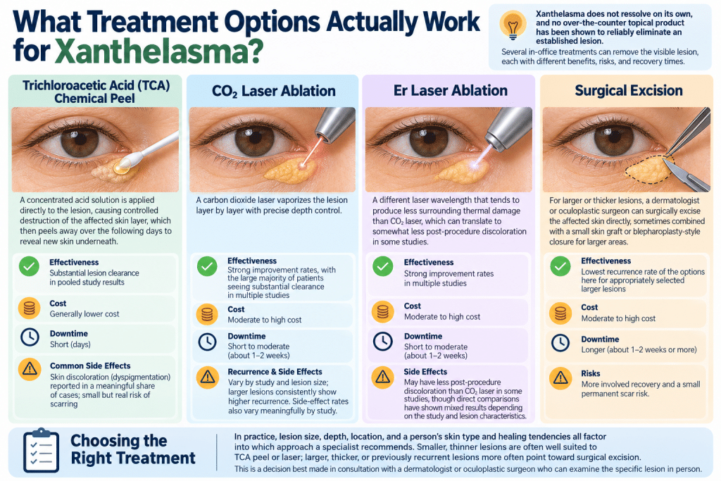
Xanthelasma does not resolve on its own, and no topical over-the-counter product has been shown to reliably eliminate an established lesion, though some approaches can help manage the underlying lipid picture (covered in Section 12). For people who want the visible lesion removed, several clinical options exist, each with a different balance of effectiveness, cost, downtime, and recurrence risk.
Trichloroacetic Acid (TCA) Chemical Peel
A concentrated acid solution is applied directly to the lesion, causing controlled destruction of the affected skin layer, which then peels away over the following days to reveal new skin underneath. This is one of the more accessible and lower-cost in-office options. Pooled results across multiple studies show substantial lesion clearance, but with a real rate of side effects: skin discoloration (dyspigmentation) has been reported in a meaningful share of cases, along with a smaller but real risk of scarring.
CO2 Laser Ablation
A carbon dioxide laser vaporizes the lesion layer by layer with precise depth control. Multiple studies report strong improvement rates, with the large majority of patients seeing substantial clearance. Recurrence and side-effect rates vary meaningfully by study and by the size of the treated lesion, with larger lesions consistently showing higher recurrence than smaller ones.
Er Laser Ablation
A different laser wavelength that tends to produce less surrounding thermal damage than CO2 laser, which can translate to somewhat less post-procedure discoloration in some studies, though direct head-to-head comparisons between the two laser types have produced mixed results depending on the specific study and lesion characteristics.
Surgical Excision
For larger or thicker lesions, particularly ones that have not responded well to peels or laser, a dermatologic or oculoplastic surgeon can surgically excise the affected skin directly, sometimes combined with a small skin graft or blepharoplasty-style closure for larger areas. This approach tends to have the lowest recurrence rate of the options here for appropriately selected larger lesions, at the cost of a more involved recovery and a small permanent scar risk.
Choosing the Right Treatment
In practice, lesion size, depth, location, and a person’s skin type and healing tendencies all factor into which approach a specialist recommends. Smaller, thinner lesions are often well suited to TCA peel or laser; larger, thicker, or previously recurrent lesions more often point toward surgical excision. This is a decision best made in consultation with a dermatologist or oculoplastic surgeon who can examine the specific lesion in person.
What Are the Recurrence Rates for Xanthelasma?
One of the least discussed but most important parts of the xanthelasma treatment conversation is recurrence. Because the underlying driver is systemic, lipid metabolism and, in many cases, oxidative stress, rather than something purely local to the treated skin, removing the visible lesion does not remove the underlying process that produced it. Recurrence is common enough that it deserves honest treatment rather than being glossed over.
Reported recurrence rates vary considerably depending on the treatment method, lesion size, and how long patients were followed after treatment. TCA peel studies have reported recurrence rates in a meaningful minority of cases, with one direct comparison study finding a notably higher recurrence rate for TCA peel than for CO2 laser treatment of similar lesions.
Laser-based approaches (CO2 and Er) have shown a wide range of recurrence rates across different studies, generally lower than TCA peel on average but still a real possibility, particularly for larger lesions. Surgical excision tends to show the lowest recurrence rates among the options discussed, though it is also the most invasive.
Across nearly every study on this topic, one pattern holds consistently: larger, thicker lesions recur at meaningfully higher rates than smaller, thinner ones, regardless of which treatment method is used. This is one of the more practical, evidence-backed reasons to address a smaller lesion earlier rather than waiting for it to grow before pursuing treatment, if treatment is something a person is considering at all.
It is also worth being direct about the underlying implication: because recurrence is tied to ongoing lipid metabolism, someone whose xanthelasma is connected to elevated LDL or ongoing oxidative stress, and who does not address those underlying factors, is more likely to see the lesion return after treatment than someone whose lipid picture has been brought under control in the meantime. This is exactly why the nutrition connection in Section 14 matters, not as an alternative to medical treatment, but as a meaningful complement to it.
Get 11 Sample Recipes from Our Bestselling Mediterranean Cookbook.
Get our 11 Sample Recipes from our Best Selling Mediterranean Protein Friendly Cookbook. These are the most delicious recipes our retreat guests love the most. Easy, simple with few ingredients per meal. Make delicious heart-friendly and healthy meals you enjoy every day.
Can At-Home and Over-the-Counter Approaches Treat Xanthelasma?
Given how often “xanthelasma removal at home” is searched, it is worth being straightforward about what home approaches can and cannot realistically achieve.
What Is Not Supported by Evidence?
No over-the-counter cream, essential oil, apple cider vinegar application, or other home remedy has been shown in credible research to reliably dissolve or remove an established xanthelasma lesion.
Anecdotal claims circulate widely online, but the biology of the lesion, lipid-laden macrophages embedded in the dermis, is not something a topical surface application is positioned to meaningfully reverse. Attempting to physically pick, scrape, or otherwise self-treat the delicate eyelid skin at home carries a real risk of infection, scarring, or damage to the eye itself and is not recommended.
What Can Help?
Addressing the underlying lipid and oxidative stress picture through diet, discussed in depth in Section 14, will not dissolve an existing lesion, but it is the one home-controllable factor with a plausible, evidence-supported mechanism for reducing the likelihood of new lesions forming and potentially supporting better outcomes after professional treatment.
This is a meaningfully different claim than “removes xanthelasma at home,” and it is the honest version of what the research supports.
The Realistic Treatment Path
For an existing visible lesion, professional in-office treatment (Section 10) is the only approach with real evidence behind it. For the broader goal of addressing what caused the lesion to form in the first place, nutrition and, where appropriate, medical management of cholesterol are the levers that matter.
What Does the Evidence Show at a Glance?
The following table summarizes the strength of the available evidence for the major claims about xanthelasma, cardiovascular risk, menopause, and treatment options.
| Claim | Evidence Strength | What the Research Shows |
|---|---|---|
| Xanthelasma predicts elevated heart attack risk independent of cholesterol | Strong | Copenhagen City Heart Study (n=12,745, 30+ year follow-up): HR 1.48 for MI, 1.39 for ischemic heart disease, adjusted for standard risk factors including cholesterol |
| Xanthelasma predicts elevated stroke risk | Weak/Not supported | Same study found no significant association with ischemic stroke or cerebrovascular disease |
| Corneal arcus predicts elevated cardiovascular risk independent of age | Weak/Mixed | Copenhagen cohort found no statistically significant association after adjustment; other smaller studies show mixed results |
| Some xanthelasma patients have entirely normal cholesterol | Strong | Multiple studies show 25 to 70 percent of xanthelasma patients are normolipidemic, with elevated lipid peroxidation markers instead |
| Menopause increases LDL and apoB | Strong | Framingham Offspring Study and related cohorts show measurable postmenopausal increases in LDL, total cholesterol, and apoB after age adjustment |
| TCA peel effectively clears lesions | Moderate | Good clearance rates reported, but with meaningful rates of dyspigmentation and higher recurrence than laser or surgery in comparative studies |
| CO2/Er laser effectively clears lesions | Moderate to Strong | Strong clearance rates reported across multiple studies; recurrence varies by lesion size |
| Surgical excision has the lowest recurrence | Moderate | Consistently reported as lowest recurrence among options, particularly for larger lesions, though most invasive |
| Home remedies remove existing xanthelasma | Not supported | No credible evidence for topical or home-based removal of established lesions |
Get 11 Sample Recipes from Our Bestselling Mediterranean Cookbook.
Get our 11 Sample Recipes from our Best Selling Mediterranean Protein Friendly Cookbook. These are the most delicious recipes our retreat guests love the most. Easy, simple with few ingredients per meal. Make delicious heart-friendly and healthy meals you enjoy every day.
How Does Nutrition Connect to Xanthelasma?
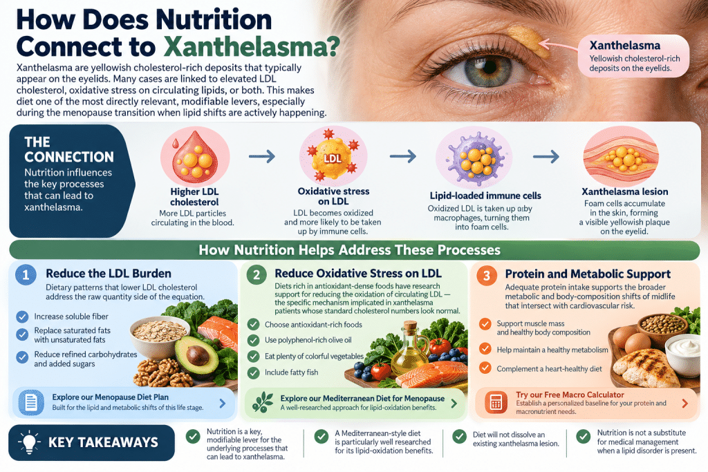
Given that a meaningful share of xanthelasma cases trace back to elevated LDL, oxidative stress on circulating lipids, or both, diet is not a peripheral detail in this conversation; it is one of the most directly relevant, modifiable levers available, particularly through the menopause transition when the underlying lipid shift described in Section 5 is actively happening.
Reducing the LDL Burden
Dietary patterns that lower LDL cholesterol, higher intake of soluble fiber, replacing saturated fat with unsaturated fat sources, and reducing refined carbohydrate and added sugar intake, address the raw quantity side of the equation. This is precisely the territory covered in our Menopause Diet Plan, built specifically around the lipid and metabolic shifts of this life stage.
Reducing Oxidative Stress on LDL
This is the piece most directly relevant to the normolipidemic xanthelasma finding in Section 4. Diets rich in antioxidant-dense foods, polyphenol-rich olive oil, colorful vegetables, and fatty fish, have research support for reducing the oxidation of circulating LDL, which is the specific mechanism implicated in xanthelasma patients whose standard cholesterol numbers look normal.
The Mediterranean dietary pattern is, not coincidentally, one of the most extensively researched dietary approaches for exactly this kind of lipid-oxidation benefit, which is why it sits at the center of our Mediterranean Diet for Menopause guide.
Protein and Metabolic Support
Adequate protein intake supports the broader metabolic and body-composition shifts of midlife that intersect with cardiovascular risk. Readers building out a full nutrition framework can start with our Free Macro Calculator to establish a personalized baseline.
None of this is a substitute for medical management when a lipid disorder is present, and none of it will dissolve an existing xanthelasma lesion once it has formed. But for the underlying process that produces the lesion in the first place, and for the broader cardiovascular risk picture the Copenhagen City Heart Study connects it to, nutrition is one of the most concretely actionable pieces of this entire topic.
What Are the Common Misconceptions About Xanthelasma?
Several common misconceptions about xanthelasma can lead to confusion about cholesterol levels, cardiovascular risk, and treatment. Understanding what the evidence shows helps separate these assumptions from the findings.
- Misconception: “If my cholesterol is normal, xanthelasma means nothing”. Not necessarily true. A substantial share of xanthelasma patients have normal standard cholesterol panels, yet still show elevated markers of lipid oxidation, a dimension of cardiovascular risk a standard panel does not measure.
- Misconception: “Xanthelasma and corneal arcus carry the same risk”. Not supported by the flagship Copenhagen City Heart Study data, which found a strong, statistically significant association for xanthelasma and heart attack risk, but not a significant one for corneal arcus after adjustment.
- Misconception: “It’s just a skin thing, purely cosmetic”. The lesion itself is harmless, but the research strongly suggests it is not “just” cosmetic in what it may indicate about underlying cardiovascular risk.
- Misconception: “Removing it fixes the underlying problem”. Removal addresses the visible lesion, not the lipid or oxidative process that produced it, which is why recurrence is common, particularly without addressing diet or, where relevant, medical cholesterol management.
- Misconception: “It only happens to people who eat poorly”. Genetics, age-related changes in LDL clearance, and the menopause-driven hormonal shift described in Section 5 all contribute independent of diet quality, and even people with excellent diets can develop xanthelasma due to genetic lipid disorders or menopausal hormone changes.
Get 11 Sample Recipes from Our Bestselling Mediterranean Cookbook.
Get our 11 Sample Recipes from our Best Selling Mediterranean Protein Friendly Cookbook. These are the most delicious recipes our retreat guests love the most. Easy, simple with few ingredients per meal. Make delicious heart-friendly and healthy meals you enjoy every day.
The Reverse Age Effect
Xanthelasma is one of the few visible signs on your face that may reflect what’s happening inside your arteries. That makes it a useful prompt for one of the most important anti-aging strategies there is: protecting your heart.
- Combine with nutrition: The lipid changes that can occur through midlife make overall dietary pattern especially important. A structured menopause diet plan can help organize day-to-day choices, while a Mediterranean-style approach emphasizes foods associated with better cardiovascular and metabolic health.
- Combine with other visible-marker awareness: Xanthelasma is not the only outward sign that can prompt a closer look at underlying health. Findings such as Frank’s sign and nail ridges can also carry associations worth understanding, even though neither should be treated as a diagnosis on its own.
- Combine with movement: Regular physical activity supports cardiovascular health, circulation, and metabolic function. Adding simple vagus nerve exercises can complement that movement routine by supporting nervous system regulation and recovery.
- Combine with sleep and stress management: Sleep quality and chronic stress both influence cardiovascular and metabolic health, so they belong in the same broader prevention framework. Building a consistent midlife sleep routine and using small, repeatable stress-management habits can help strengthen those foundations over time.
Get 11 Sample Recipes from Our Bestselling Mediterranean Cookbook.
Get our 11 Sample Recipes from our Best Selling Mediterranean Protein Friendly Cookbook. These are the most delicious recipes our retreat guests love the most. Easy, simple with few ingredients per meal. Make delicious heart-friendly and healthy meals you enjoy every day.
Frequently Asked Questions
What is xanthelasma?
Xanthelasma is a soft, flat or slightly raised, yellowish patch that forms on or near the eyelids, usually at the inner corners closest to the nose. It is made up of lipid-laden immune cells (foam cells) that accumulate in the skin’s dermis layer and is generally harmless on its own, though it can indicate underlying lipid or cardiovascular issues.
Is xanthelasma always caused by high cholesterol?
No. While elevated LDL cholesterol is a common contributor, research shows that roughly a quarter to as much as seventy percent of xanthelasma patients across different studies have entirely normal standard cholesterol panels. In these cases, elevated markers of lipid oxidation appear to play a role instead.
Does xanthelasma mean I’m going to have a heart attack?
No. The Copenhagen City Heart Study found xanthelasma associated with a roughly 48 percent higher relative risk of heart attack over decades of follow-up, a real and statistically significant finding, but this reflects a population-level association, not an individual prediction or a guarantee.
Why does xanthelasma often appear after menopause?
Estrogen supports the efficiency of LDL receptors, which clear cholesterol from the bloodstream. As estrogen declines through perimenopause and menopause, LDL clearance becomes less efficient, and LDL cholesterol, total cholesterol, and apolipoprotein B levels rise measurably, creating conditions more favorable to xanthelasma formation.
Can xanthelasma go away on its own?
No. Unlike temporary skin irritation, xanthelasma is a structural accumulation of lipid-laden cells in the dermis and does not resolve without treatment. It can, however, slowly grow larger if left untreated.
What is the difference between xanthelasma and corneal arcus?
Xanthelasma is a yellow patch on the eyelid skin itself. Corneal arcus is a grayish-white ring around the edge of the cornea, the clear surface of the eye. Both involve lipid deposition, but they occur in different tissues, and the evidence connecting them to cardiovascular risk differs, xanthelasma has stronger, more specific supporting evidence than corneal arcus in the largest study on the topic.
Can I remove xanthelasma at home?
No topical or home remedy has credible evidence for removing an established xanthelasma lesion, and attempting to pick or scrape the delicate eyelid skin risks infection and scarring. Professional treatment options (TCA peel, laser, or surgical excision) are the only approaches with meaningful evidence behind them.
What treatment has the lowest recurrence rate?
Across the available studies, surgical excision tends to show the lowest recurrence rates, particularly for larger lesions, though it is also the most invasive option. Laser treatments (CO2 and Er) generally show lower recurrence than TCA chemical peel, though results vary by study and lesion size.
Will treating xanthelasma prevent it from coming back?
Not necessarily. Because the underlying driver is systemic lipid metabolism rather than something purely local to the skin, removing the visible lesion does not address the process that produced it. Addressing diet and, where relevant, medical cholesterol management alongside treatment gives the best chance of reducing recurrence.
Should I get my cholesterol checked if I notice xanthelasma?
Yes, this is one of the most useful, low-stakes actions to take. A full lipid panel, not just total cholesterol, gives you and your physician the clearest picture of whether elevated LDL is contributing, and is worth doing even if you suspect your cholesterol is normal, given how common normolipidemic xanthelasma is.
Is xanthelasma more common in women?
It affects both sexes, but the sharp rise in prevalence tied to the estrogen-driven LDL changes of the menopause transition makes midlife women a population where it shows up with particular frequency, often for the first time in their late forties or fifties.
Does insurance cover xanthelasma removal?
This varies by insurer and is frequently classified as a cosmetic procedure rather than a medically necessary one, which can affect coverage. It is worth confirming directly with your insurance provider and the treating specialist before scheduling a procedure.
Can xanthelasma come back after successful removal?
Yes, recurrence is common enough to be a genuine consideration, not a rare exception, particularly for larger lesions and particularly when the underlying lipid or oxidative stress picture is not addressed alongside treatment.
Is corneal arcus dangerous?
Based on the largest and most rigorously controlled study on the topic, corneal arcus was not found to independently predict cardiovascular risk once age and standard risk factors were accounted for, unlike xanthelasma. It remains a topic with more mixed evidence overall and is worth exploring in its own dedicated discussion.
I write 3 essays per week on Lifestyle Medicine for women in midlife on what is happening in the body and what to do about it. No fluff, no hype. Free Subscription.
References
- Christoffersen, M., Frikke-Schmidt, R., Schnohr, P., Jensen, G. B., Nordestgaard, B. G., & Tybjærg-Hansen, A. (2011). Xanthelasmata, arcus corneae, and ischaemic vascular disease and death in general population: prospective cohort study. BMJ, 343, d5497.
- Schaefer, E. J., Lamon-Fava, S., Cohn, S. D., Schaefer, M. M., Ordovas, J. M., Castelli, W. P., & Wilson, P. W. (1994). Effects of age, gender, and menopausal status on plasma low density lipoprotein cholesterol and apolipoprotein B levels in the Framingham Offspring Study. Journal of Lipid Research, 35(5), 779-792.
- Bergman, R. (1994). The pathogenesis and clinical significance of xanthelasma palpebrarum. Journal of the American Academy of Dermatology, 30(2), 236-242.
- Dey, A., Aggarwal, R., & Dwivedi, S. (2013). Cardiovascular profile of xanthelasma palpebrarum. BioMed Research International, 2013, 932138.
- Nassar, G. T., & Krishan, K. (2023). Xanthelasma palpebrarum. StatPearls, National Library of Medicine.
- Rohrich, R. J., Janis, J. E., & Pownell, P. H. (2002). Xanthelasma palpebrarum: a review and current management principles. Plastic and Reconstructive Surgery, 110(5), 1310-1314.
- Laftah, Z., & Al-Niaimi, F. (2013). Xanthelasma: an update on treatment modalities. Journal of Cutaneous and Aesthetic Surgery, 6(4), 203-207.
- Christoffersen, M., Frikke-Schmidt, R., Schnohr, P., Jensen, G. B., Nordestgaard, B. G., & Tybjærg-Hansen, A. (2014). Visible age-related signs and risk of ischemic heart disease in the general population: A prospective cohort study. Circulation, 129(9), 990-998.
- American Heart Association. (2023). Heart disease and stroke statistics update. Circulation, 147(8), e93-e621.
Disclaimer: This article is for educational purposes only and does not constitute medical advice, diagnosis, or treatment. Xanthelasma may be associated with cholesterol abnormalities and increased cardiovascular risk, but its presence alone does not indicate heart disease. If you notice yellow patches around your eyelids, consult a qualified healthcare provider for proper evaluation and cholesterol testing. Avoid attempting at-home removal, as the delicate eyelid area is vulnerable to irritation, scarring, and injury. Treatment decisions and dietary changes should be discussed with a healthcare professional based on your individual health needs.
Get 11 Sample Recipes from Our Bestselling Mediterranean Cookbook.
Get our 11 Sample Recipes from our Best Selling Mediterranean Protein Friendly Cookbook. These are the most delicious recipes our retreat guests love the most. Easy, simple with few ingredients per meal. Make delicious heart-friendly and healthy meals you enjoy every day.

Terry Tateossian is a Menopause Lifestyle Medicine Coach, Certified Personal Trainer & Nutritionist and the Founder of THOR: The House of Rose, a wellness brand serving women over 40 through retreats, coaching, macro-nutrition and community. As 25-year founder and entrepreneur, Terry spent two and a half decades building and running successful start-up businesses, an experience that put her on the front line of founder burnout long before she could name it. After facing serious health challenges, early onset menopause, and emotional eating while running her agency and raising two children, Terry rebuilt her health in her 40s and lost more than 80 pounds through evidence-based nutrition, training, and mindset work. Today, she helps women get strong, improve confidence, support hormone health, and create a stronger second half of life. Terry has been featured in major media outlets and is available for podcasts, expert commentary, brand collaborations, and speaking engagements on midlife health, reinvention, emotional eating, menopause wellness, and strength training for longevity. Get her free macro calculator (her cookbook companion) to start your journey to back to health.